")
What is Electroencephalogram (EEG)?
It is a Window into Your Brain and Objective Assessment of Neural & Mental capabilities
EEG is fast, noninvasive, painless and precise measure of brain functional activity. The intimate, dynamic structure of this activity is information-rich about the underlying cellular and intercellular processing, brain physiological, cognitive and mental functions and resources, brain states and conditions. EEG, from one hand, reflects functional brain state which comprises dynamical processes, and from another hand, EEG may allow to modify the functional architecture of the cortex thus affecting brain and body activity.
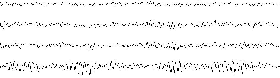
Digital EEG is the digitally recorded electrical activity generated by the brain. In general, EEG is obtained using electrodes placed on the scalp according to an International 10-20 System with a conductive gel. In the brain, there are millions of neurons, each of which generates small electric voltage fields. The aggregate of these electric voltage fields creates an electrical reading which electrodes on the scalp are able detect and record. Therefore, EEG is the superposition of many simpler signals.
 The core principle of quantitative EEG (QEEG) lies in "precise and objective quantification" of electrophysiological brain activity by computerized analytic and statistical techniques. It permits to extract more precise information from the brain electrical activity than when using the conventional simple un-aided visual inspection of EEG signal. QEEG evaluates the manner in which a particular person’s brain physiological, cognitive and mental functions organized and how brain uses its strength and resources to cope with stress or disease and compensate it.
Numerous studies have repeatedly demonstrated the high stability, reliability and specificity of certain QEEG parameters. These parameters were being studied quantitatively in large samples of healthy functioning individuals across different nations and wide age ranges. Numerous studies confirmed the high specificity of normative distributions of QEEG parameters within different EEG frequencies (delta, theta, alpha, and beta bands). Positive findings different from the normative databases in healthy, normally functioning individuals have repeatedly been shown to be within the chance levels, with very high test-retest reliability. Normative data have been extended to cover the age range from 1 to 95 years for each EEG electrode in the standardized International 10-20 System and include many parameters of QEEG. The independence of the normative QEEG descriptors from cultural and ethnic factors enables objective assessment of brain integrity in individuals of any age, origin or background. Naturally, the statistically significant deviation of the QEEG parameters from normative levels indicates the dysfunctions and abnormalities associated with neurological, developmental, and psychiatric disorders. Numerous studies support this claim.
There are several reasons why EEG is an exceptional tool for studying the neurocognitive processes underlying human behavior and cognition (Cohen (2011). It’s about time. Frontiers in Human Neuroscience, 5(2), 1–15):
EEG has a very high time resolution and captures cognitive processes in the time frame in which cognition naturally occurs. It captures the physiological changes underlying the cognitive processes much better than other brain imaging techniques (such as MRI or PET scanners).
EEG directly measures neural activity. Techniques like MRI only measure neural activity indirectly and lacking a deeper understanding of the relationship between what is measured and how it relates to cognitive processing.
EEG monitors cognitive-affective processing in absence of behavioral responses. Brain processes ultimately drive behavior. However, if you’re interested in mental processes such as response inhibition, creativity or meditation, the behavioral effects might be very subtle. By contrast, these processes are ideal candidates for EEG as they are accompanied by distinguishable electrical brain activation patterns.
EEG is absolutely non-invasive and non-stressful procedure. Other techniques (such as MRI or PET scanners) are minimally invasive or invasive and stressful.
Selected references
-
Basar E. Brain function and oscillations: I. Brain oscillations, principles and approaches. Berlin: Springer; 1998
-
Fingelkurts Al.A., Fingelkurts An.A. Short-term EEG spectral pattern as a single event in EEG phenomenology. The Open Neuroimaging Journal, 2010;4:130-156
-
Fingelkurts Al.A., Fingelkurts An.A., Ermolaev V.A., Kaplan A.Ya. Stability, reliability and consistency of the compositions of brain oscillations. International Journal of Psychophysiology, 2006;59(2):116-126
-
Fingelkurts An.A., Fingelkurts Al.A. Alpha rhythm Operational Architectonics in the continuum of normal and pathological brain states: Current state of research. International Journal of Psychophysiology, 2010;76(2):93-106
-
Fingelkurts An.A., Fingelkurts Al.A. Brain-mind Operational Architectonics imaging: technical and methodological aspects. The Open Neuroimaging Journal, 2008;2:73-93
-
Fingelkurts An.A., Fingelkurts Al.A. Making complexity simpler: Multivariability and metastability in the brain. International Journal of Neuroscience, 2004;114(7):843-862
-
Fingelkurts An.A., Fingelkurts Al.A. Timing in cognition and EEG brain dynamics: discreteness versus continuity. Cognitive Processing, 2006;7(3):135-162
-
Gevins A. Electrophysiological Imaging of Brain Function. P. 175–188. In: Brain mapping. The methods. Toga AW, Mazzoitta JC (eds). 2 edition. Elsevier Science (USA), 2002; pp. 877.
-
Gevins A. The future of electroencephalography in assessing neurocognitive functioning. Electroencephalogr. Clinical Neurophysiology, 1998;106:165–172
-
Hughes J.R., John E.R. Conventional and Quantitative Electroencephalography in Psychiatry. The Journal of Neuropsychiatry and Clinical Neurosciences, 1999;11:190-208
-
Inui K., Motomura E., Okushima R., et al: Electroencephalographic findings in patients with DSM-IV mood disorder, schizophrenia, and other psychotic disorders. Biological Psychiatry, 1998;43:69–75
-
John E.R., Karmel B.Z., Corning W.C., Easton P., Brown D., Ahn H., John M., Harmony T., Prichep L., Toro A., Gerson I., Bartlett F., Thatcher R., Kaye H., Valdes P., Schwartz E. Neurometrics: Numerical taxonomy identifies different profiles of brain functions within groups of behaviourally similar people. 1977;196(4297):1393-1410
-
John E.R., Prichep L.S., Winterer G., Herrmann W.M., diMichele F., Halper J., Bolwig T.G., Cancro R. Electrophysiological subtypes of psychotic states. Acta Psychiatr Scand 2007;116:17–35
-
Johnstone J, Gunkelman J, Lunt J. Clinical database development: characterization of EEG phenotypes. Clinical EEG and Neuroscience, 2005;36(2):99-107
-
Kanda P.A.dM., Anghinah R., Smidth M.T., Silva J.M. The clinical use of quantitative EEG in cognitive disorders. Dementia & Neuropsychologia, 2009 ;3(3):195-203
-
Knyazev G.G., Savostyanov A.N., Levin E.A. Uncertainty, anxiety, and brain oscillations. Neuroscience Letters, 2005;387:121–125
-
Nunez P.L. Physiological Foundations of Quantitative EEG Analysis. In: Shanbao Tong and Nitish V. Thakor, editors. Quantitative EEG Analysis Methods and Clinical Applications. ARTECH HOUSE, 2009, p.1-22
-
Prichep L.S., John E.R., Ferris S.H., Rausch L., Fang Z., Cancro R., Torossian C., Reisberg B. Prediction of longitudinal cognitive decline in normal elderly with subjective complaints using electrophysiological imaging. Neurobiology of Aging, 2006;27:471–481
-
Sanei S., Chambers J.A. EEG signal processing. John Wiley & Sons Ltd, 2007, pp.289
-
Schutter D.J.L.G., Leitner C., Kenemans J.L., van Honk J. Electrophysiological correlates of cortico-subcortical interaction: A cross-frequency spectral EEG analysis. Clinical Neurophysiology, 2006;117:381–387
-
Shelley B.P., Trimble M.R., Boutros N.N. Electroencephalographic cerebral dysrhythmic abnormalities in the trinity of nonepileptic general population, neuropsychiatric, and neurobehavioral Disorders. The Journal of Neuropsychiatry and Clinical Neurosciences, 2008;20:7–22
-
Thatcher R.W., Lubar J.F. History of the scientific standards of QEEG normative databases. Introduction to QEEG and Neurofeedback: Advanced Theory and Applications” Thomas Budzinsky, H. Budzinski, J. Evans and A. Abarbanel editors, Academic Press, San Diego, Calif (2008)